

Knee Overview
Knee Anatomy
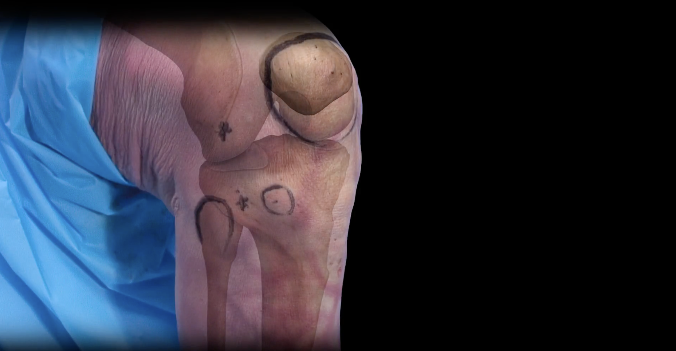
The knee joint is one of the most complex joint in the human body. The knee is designed to handle load, large amounts of stress and the demands placed on it every day. The knee can bend, twist, flex and offer power and strength for daily activities or sports. The anatomy of the knee and its flexibility can make it vulnerable to injury. Orthopedic Surgeon and Sports Medicine specialist, Dr. Jonathan Koscso, successfully diagnoses and treats patients in Sarasota, FL and the surrounding Gulf Coast region who have experienced a knee injury.

Bony Anatomy of the Knee
The knee consists of three main bones:
Femur: Also called the thigh bone
Tibia: Also called the shin bone
Patella: Also called the kneecap
The Fibula is a small bone in the lower leg that runs alongside the lateral (outer) aspect of the tibia. The fibula carries only minimal load (weight) of the body. It extends past the lower end of the tibia and forms the outer part of the ankle providing stability to this joint.
Ligaments of the Knee
The anatomy of the knee consists of several different ligaments that act to support and stabilize the knee throughout its range of motion. These ligaments are:
ACL: Anterior Cruciate Ligament, this ligament is the most commonly injured ligament in the knee requiring surgery. The ACL functions to keep the tibia from sliding forward relative to the femur. The ACL is located in the middle of the knee, connecting the front of the tibia to the back of the femur.
PCL: Posterior Cruciate Ligament, this ligament is the mirror ligament to the ACL. Along with the ACL ligament, the PCL forms an “X” in the center of the knee. The PCL connects the back of the tibia to the front of the femur, preventing the femur from sliding forward on the tibia and visa-versa. The PCL is the largest and strongest ligament in the knee, making it less susceptible to injury than the ACL.
MCL: Medial Collateral Ligament, is a very commonly injured ligament in the knee. The MCL runs down the medial (inner) part of the knee and is responsible for side-to-side stability. The MCL keeps the knee from collapsing inward.
LCL: Lateral Collateral Ligament, this ligament runs down the outside of the knee and is also responsible for supporting side-to-side movement. The LCL keeps the knee from collapsing outward.
MPFL: Medial Patello-Femoral Ligament, this ligament connects the medial (inner) side of the patella to the medial side of the femur bone and is an important stabilizer of the patella throughout knee range of motion. Injury to the MPFL from a patellar dislocation can lead to ongoing patellar instability.

Soft Tissue Structures of the Knee
Supporting structures within the anatomy of the knee help keep the knee strong, functional, and able to perform a wide range of motion. Important structures of the knee include:
Cartilage: The knee has two types of cartilage that make up the joint:
Articular cartilage: Covers the ends of the bones and allows the smooth, pain-free glide of the bones in the knee. Damage to the cartilage can come in a couple different varieties. Focal articular cartilage defects occur typically in athletes with traumatic injuries and can be treated with various cartilage repair/restoration techniques. Knee arthritis, on the other hand, is usually due to chronic breakdown of the articular cartilage in the knee.
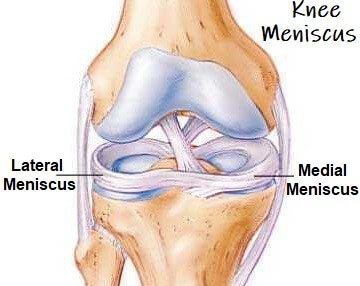
Meniscus: Each knee has two crescent-shaped discs of cartilage that act as shock absorbers in the knee. The meniscus plays a vital role in the health of the knee, allowing proper weight distribution and stability. The job of the meniscus is to keep the bones from rubbing together while walking, running, flexing, or extending the knee. Meniscus tears can lead to breakdown of the articular cartilage of the knee due loss of this shock absorbing effect.
Tendons: The muscles are attached to the bones by strong tissues called tendons. Tendons connect muscle to bone, whereas ligaments connect bone to bone. Two important tendons in the knee are:
Patellar Tendon: connects the patella to the tibia
Quadriceps Tendon: connects the quadriceps muscles to the patella at the front of the thigh.
Muscles: The strength of the knee comes from the surrounding muscles which also offer flexibility.
Hamstrings: Three muscles in the back of the leg that bend the knee.
Quadriceps: Four muscles in the front of the knee that straighten the knee.
Gluteal Muscles: While not technically part of the muscle groups within the knee, the gluteal muscles are important for proper knee positioning. The gluteal muscles, or “glutes” are made up of the gluteus maximus, medius, and minimus.
Bursae: Fluid-filled sacs within the knee that reduce friction and help prevent pain and inflammation.

About the Author

Dr. Jonathan Koscso is an orthopedic surgeon and sports medicine specialist at Kennedy-White Orthopaedic Center in Sarasota, FL. Dr. Koscso treats a vast spectrum of sports conditions, including shoulder, elbow, knee, and ankle disorders. Dr. Koscso was educated at the University of South Florida and the USF Morsani College of Medicine, followed by orthopedic surgery residency at Washington University in St. Louis/Barnes-Jewish Hospital and sports medicine & shoulder surgery fellowship at the Hospital for Special Surgery in New York City, the consistent #1 orthopaedic hospital as ranked by U.S. News & World Report. He has been a team physician for the New York Mets, Iona College Athletics, and NYC’s PSAL.
Disclaimer: All materials presented on this website are the opinions of Dr. Jonathan Koscso and any guest writers, and should not be construed as medical advice. Each patient’s specific condition is different, and a comprehensive medical assessment requires a full medical history, physical exam, and review of diagnostic imaging. If you would like to seek the opinion of Dr. Jonathan Koscso for your specific case, we recommend contacting our office to make an appointment.