

Shoulder Overview
Shoulder Anatomy
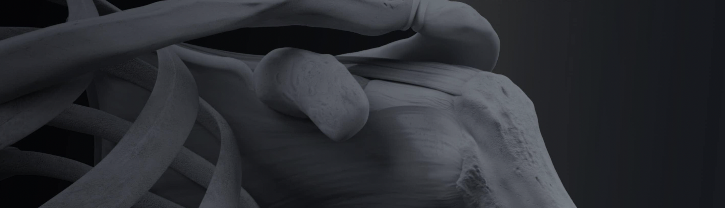
The shoulder joint is the largest, most flexible, and one of the most complex joints in the human body. The shoulder is made up of three bones: the humerus (upper arm bone), the clavicle (collar bone) and the scapula (shoulder blade). Also within the shoulder are three small joints that work together, giving the shoulder a large range of mobility and motion. These joints in the shoulder are the acromioclavicular joint, the sternoclavicular joint and the glenohumeral joint. The joint known as the “ball and socket” joint is the glenohumeral joint. Because of the complexity of the shoulder, its flexibility and the range of motion make it inherently susceptible to injury. Shoulder pain can be common and can stem from a number of different conditions.
Orthopedic Surgeon and Sports Medicine specialist, Dr. Jonathan Koscso, successfully diagnoses and treats patients in Sarasota, FL and the surrounding Gulf Coast region who have experienced a shoulder injury.

Bony Anatomy of the Shoulder
Three major bones meet to form the shoulder joint, each playing a critical role in the function of the shoulder:
Clavicle: Also called the collarbone, this thin, flat bone stabilizes movement in the shoulder. The clavicle is found at the front of the shoulder, extending from the center of the body (sternum) to the end of the shoulder.
Humerus: The ball of the upper arm bone that connects the scapula and the clavicle. The humerus fits into the scapula (glenoid) forming the ball and socket joint.
Scapula: Also called the shoulder blade, this flat, triangular bone is found at the back of the shoulder. The scapula connects the clavicle to the acromioclavicular joint.
Soft Tissue Anatomy of the Shoulder
The shoulder cannot offer mobility and strength without other supporting structures that help it move and keep it functioning properly. Shoulder stability is achieved with the help of muscles, tendons, ligaments and cartilage. These important structures include:
Cartilage: Often called articular cartilage, this slippery tissue covers both the ends of the bones and allows them to glide together easily. In the glenohumeral joint, cartilage covers both the humeral head and the glenoid socket. The breakdown of this cartilage can cause shoulder arthritis.
Labrum: A strong rubber-like ring of cartilage attached to the rim of the shoulder socket which holds the ball of the humerus firmly within the glenoid socket. Damage to the labrum can cause shoulder instability and frequent dislocations. Along the top of the glenoid socket, the long head of the biceps tendon attaches to the superior labrum, and pathologic conditions at this insertion can cause a SLAP tear (superior labrum, anterior to posterior). Additionally, the biceps tendon itself can become inflamed and lead to significant pain in the front of the shoulder, a condition called biceps tendinitis.
Bursae: Located between the bone and the surrounding soft tissue, these small sacs of fluid lubricate and cushion the rotator cuff. In pathologic subacromial impingement, this tissue can become inflamed and painful when reaching overhead.
Rotator Cuff: A grouping of 4 muscles and tendons that surround the shoulder joint. They help hold the ball of the humerus in place within the socket of the glenoid. These muscles and tendons also provide the motor function necessary to move the arm. Thus, the rotator cuff is responsible for the strength of the shoulder, as well as its stability. With a tear in the rotator cuff, using the arm can be painful and quite limited due to weakness. Over time, a large chronic rotator cuff tear can advance to include cartilage breakdown in the shoulder joint, a condition known as cuff tear arthropathy (a variant of shoulder arthritis).


About the Author

Dr. Jonathan Koscso is an orthopedic surgeon and sports medicine specialist at Kennedy-White Orthopaedic Center in Sarasota, FL. Dr. Koscso treats a vast spectrum of sports conditions, including shoulder, elbow, knee, and ankle disorders. Dr. Koscso was educated at the University of South Florida and the USF Morsani College of Medicine, followed by orthopedic surgery residency at Washington University in St. Louis/Barnes-Jewish Hospital and sports medicine & shoulder surgery fellowship at the Hospital for Special Surgery in New York City, the consistent #1 orthopaedic hospital as ranked by U.S. News & World Report. He has been a team physician for the New York Mets, Iona College Athletics, and NYC’s PSAL.
Disclaimer: All materials presented on this website are the opinions of Dr. Jonathan Koscso and any guest writers, and should not be construed as medical advice. Each patient’s specific condition is different, and a comprehensive medical assessment requires a full medical history, physical exam, and review of diagnostic imaging. If you would like to seek the opinion of Dr. Jonathan Koscso for your specific case, we recommend contacting our office to make an appointment.